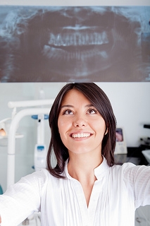
X-rays have been used for a long time in dentistry and orthodontics, and with advancements in technology, the level of radiation has reduced considerably. Although risk exists in any health diagnostic operation, the benefits must outweigh the risks. Indeed, dental X-rays fall under this group.
To create an image, X-rays are applied to a form of film. The number of X-rays needed to create this picture varies with the velocity of the film. Speed E or F is highly recommended, and up to 50 percent less than speed E or F film is needed for digital X-rays. To generate a quality image, the digital X-ray program will change the exposure. Digital X-rays are becoming the latest and most prevalent norm.
Lead aprons have been used to decrease the amount of radiation dispersion. In order to concentrate the X-ray beam, all X-ray units have a cone, so the exposure is highly localized. Although lead aprons and thyroid collars are worn during procedures, out of precaution we ask our pregnant patients to avoid orthodontic x-ray exams until after their pregnancy.
As well as healthcare testing and treatment methods, we get radiation exposure from environmental causes. In order to put this in context, a person is supposed to have 360mRem from the sun, air, etc. per year in one year. For contrast, a single range of X-rays for bitewing is 0.3mRem. Radiation will accumulate over a lifetime in our body, and if possible, further exposure should be avoided.
